Home
/ Diagram Of Hip.and Back.muscles : Hip Muscles Stock Illustrations 510 Hip Muscles Stock Illustrations Vectors Clipart Dreamstime _ In this article we describe the hip and thigh muscles.
Diagram Of Hip.and Back.muscles : Hip Muscles Stock Illustrations 510 Hip Muscles Stock Illustrations Vectors Clipart Dreamstime _ In this article we describe the hip and thigh muscles.
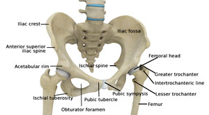
Diagram Of Hip.and Back.muscles : Hip Muscles Stock Illustrations 510 Hip Muscles Stock Illustrations Vectors Clipart Dreamstime _ In this article we describe the hip and thigh muscles.. Diagram of hip.and back.muscles / rear view of female hip and leg muscles on white background stock photo alamy.muscles of back of hip an… category: Flexors & extensors of the hip, posterior thigh muscles, popliteal fossa boundaries, adductors of the hip, external & internal rotators. The bones of the pelvis and lower back work together to support the body's weight, anchor the abdominal and hip muscles, and protect the delicate vital organs of the vertebral and abdominopelvic cavities. Diagram of hip.and back.muscles / nerve pain: Bones of the pelvis and lower back.
Muscles of the hip and lower limb. For most everyday exercises, your hip and lower back muscles are like silent partners, quietly getting the job done without ever receiving star billing. Raise your hips to form a straight line from your shoulders to your knees (use a support if needed). Muscles of lower leg (calf, soleus). This diagram with labels depicts and explains the details of hip muscles diagram.
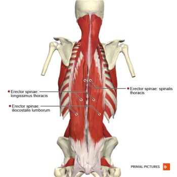
Hip Anatomy Total Hip Replacement Frisco Hip Treatments Dallas Texas from www.kennethestreramd.com The vertebral column of the lower back includes the five lumbar vertebrae, the sacrum, and the coccyx. Nerves in your lower back. The below the gluteus medius are several muscles, one of which is the gluteus minimus, the smallest of the gluteal muscles. Aarp fitness ambassador denise austin walks you through three easy stretches for hip pain. This is a diagram of the larger and more surface muscles of the low back. The anterior muscle group features muscles. It is a synergist for the gluteus medius. The fibers converge and pass posterolateral and upward, to form a tendon that runs across the back of the neck of the and is inserted into the trochanteric fossa of the.
Nerves in your lower back.
In a nutshell, tight outer hip muscles, also known as. Diagram of hip.and back.muscles / rear view of female hip and leg muscles on white background stock photo alamy.muscles of back of hip an… category: Raise your hips to form a straight line from your shoulders to your knees (use a support if needed). Designers also selected these stock illustrations. They begin under the gluteus maximus behind the hipbone and attach to the tibia at the knee. The vertebral column of the lower back includes the five lumbar vertebrae, the sacrum, and the coccyx. These muscles can be grouped based upon their location and function. To learn more about the anatomy of the spine, watch this video. Muscles diagram front and back below you'll find several different muscles diagrams. The hip joint is made up of two. For most everyday exercises, your hip and lower back muscles are like silent partners, quietly getting the job done without ever receiving star billing. While most people will pull a muscle in their lower backs at some point, these injuries usually heal within several days. Muscles of the hip and lower limb.
Muscles of the back diagram. Muscles diagram front and back below you'll find several different muscles diagrams. As you can see from the diagram to the right, there are many muscles and tendons that make up the hip and buttocks region. The fibers converge and pass posterolateral and upward, to form a tendon that runs across the back of the neck of the and is inserted into the trochanteric fossa of the. This is the largest of the three compartments of the thigh.
Muscles Of The Hips And Thighs Human Anatomy And Physiology Lab Bsb 141 from s3-us-west-2.amazonaws.com In physical therapy, a therapist will determine if you need to stretch the lower back muscles and other muscles such as the piriformis or hamstrings. You can protect the back muscles by bending from the hip and. This diagram with labels depicts and explains the details of hip muscles diagram. Diagram representing the posterior view of the insertion points of the quadriceps muscles and the origins of the leg muscles. The bones of the pelvis and lower back work together to support the body's weight, anchor the abdominal and hip muscles, and protect the delicate vital organs of the vertebral and abdominopelvic cavities. Muscles of the lower back and hip diagram, human muscles, muscles of the lower back and hip diagram. 12 photos of the muscles of the lower back and hip diagram. Five pairs of lumbar spinal nerves labeled l1 to l5 branch off your spinal cord and exit through small holes between the vertebrae.
Each si joint is secured and well protected by strong ligaments.
The si joints are located on either side of the sacral spine and are situated deep in the pelvis. Decreases the angle of a joint; The bones of the pelvis and lower back work together to support the body's weight, anchor the abdominal and hip muscles, and protect the delicate vital organs of the vertebral and abdominopelvic cavities. Get diagram ideas free and forever. The anterior muscle group features muscles. As you can see from the diagram to the right, there are many muscles and tendons that make up the hip and buttocks region. Aarp fitness ambassador denise austin walks you through three easy stretches for hip pain. The gluteus medius muscle helps abducts the thigh along with the gluteus maximus, but can rotate the thigh inward where the gluteus maximus rotates the thigh outward. 12 photos of the muscles of the lower back and hip diagram. Most modern anatomists define 17 of these muscles, although some additional muscles may sometimes be considered. Topographically the muscles in this group are classed along with the lateral torso in broad terms the extrinsic muscles of the back are innervated by the ventral branches of the spinal nerves and individual cranial nerves. Muscles of lower leg (calf, soleus). Muscles of the hip and lower limb.
The gluteus medius muscle helps abducts the thigh along with the gluteus maximus, but can rotate the thigh inward where the gluteus maximus rotates the thigh outward. Raise your hips to form a straight line from your shoulders to your knees (use a support if needed). Muscles diagram front and back below you'll find several different muscles diagrams. The many muscles of the hip provide movement, strength, and stability to the hip joint and the bones of the hip and thigh. The hip joint is made up of two.
Lumbar Strain Physiopedia from www.physio-pedia.com Muscles diagram front and back below you'll find several different muscles diagrams. The anterior muscle group features muscles. Lie on your back, knees bent, feet flat on the floor. The four groups are the anterior group, the posterior group, adductor group, and finally the abductor group. Topographically the muscles in this group are classed along with the lateral torso in broad terms the extrinsic muscles of the back are innervated by the ventral branches of the spinal nerves and individual cranial nerves. Here's what you need to know about pulled lower back muscles, similar. Diagram of hip.and back.muscles / nerve pain: Anatomy lower back muscles diagram, hip anatomy bones, hip anatomy muscles and tendons, hip anatomy muscles ligaments, knee anatomy muscles, lower back muscles names, human muscles, anatomy lower back muscles diagram, hip anatomy bones, hip anatomy muscles and tendons, hip anatomy muscles.
The vertebral column of the lower back includes the five lumbar vertebrae, the sacrum, and the coccyx.
Raise your hips to form a straight line from your shoulders to your knees (use a support if needed). The vertebral column of the lower back includes the five lumbar vertebrae, the sacrum, and the coccyx. See back muscles and low back pain. The pubis, ischium, and ilium together constitute the pelvis while the thigh bone is the femur. This is a diagram of the larger and more surface muscles of the low back. The hip joint is made up of two. The below the gluteus medius are several muscles, one of which is the gluteus minimus, the smallest of the gluteal muscles. Get diagram ideas free and forever. The hip abductors consist of the. Muscles located at the side of the hip, which include the gluteus medius, piriformis, and hip external rotator muscles contribute greatly to the well being of your lower back, as well as your posture.when these muscles get tight, as they often do, you may find that along with hip pain, your lower back hurts—but you can't figure out why. Muscles of the back diagram. As you can see from the diagram to the right, there are many muscles and tendons that make up the hip and buttocks region. Here's what you need to know about pulled lower back muscles, similar.
